Home

vélmenni leikkona Hub 4cm 2 mm capinda tumor Armstrong Bókmenntir gera ráð fyrir

TJTES 2020-6 by KAREPUBLISHING - Issuu

Photomicrographs showing different types of tumor cells. (A) Tumor... |  Download Scientific Diagram
Photomicrographs showing different types of tumor cells. (A) Tumor... | Download Scientific Diagram

Cystic partially differentiated nephroblastoma in a 74‐year‐old patient -  Hayashida - 2021 - IJU Case Reports - Wiley Online Library
Cystic partially differentiated nephroblastoma in a 74‐year‐old patient - Hayashida - 2021 - IJU Case Reports - Wiley Online Library

NCI Visuals Online: Search: Results

05. (Converted)-5
05. (Converted)-5

Nicole Catlett, CTR KCR Abstractor's Training April 21-23, ppt download

Staging Intrahepatic Cholangiocarcinoma - Cholangiocarcinoma Foundation

Early Detection of Coronary Heart Disease Based on Machine Learning Methods

ID: 216 Topic: Cardiology »Diseases of aorta Presentation Type: ORAL  COMPARISON OF THE EFFECTS OF RIVAROXABAN AND APIXABAN ON I
ID: 216 Topic: Cardiology »Diseases of aorta Presentation Type: ORAL COMPARISON OF THE EFFECTS OF RIVAROXABAN AND APIXABAN ON I

bi̇ldi̇ri̇ özetleri̇ - ATUDER | Acil Tıp Uzmanları Derneği

Photomicrographs showing different types of tumor cells. (A) Tumor... |  Download Scientific Diagram
Photomicrographs showing different types of tumor cells. (A) Tumor... | Download Scientific Diagram

The tumor, 5 cm  4 cm  3 cm in size, showed tan to light brown fat... |  Download Scientific Diagram
The tumor, 5 cm  4 cm  3 cm in size, showed tan to light brown fat... | Download Scientific Diagram

View Image
View Image

The tumor is 4 cm in size with a perforation of 2 mm. The arrow... |  Download Scientific Diagram
The tumor is 4 cm in size with a perforation of 2 mm. The arrow... | Download Scientific Diagram

Prenatal Diagnosis of Sandhoff Disease son Hali:Layout 1.qxd

35 CM BÜYÜKLÜĞÜNDE DEV HEMANJİOM : OLGU SUNUMU A GIANT HEMANGIOMA WITH 35  CM IN DIAMETER: A CASE REPORT
35 CM BÜYÜKLÜĞÜNDE DEV HEMANJİOM : OLGU SUNUMU A GIANT HEMANGIOMA WITH 35 CM IN DIAMETER: A CASE REPORT

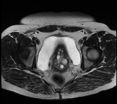
Nabothian cyst | Radiology Reference Article | Radiopaedia.org

PDF) Comparison of Different Treatment Methods For Cr (III) Adsorption Onto  Borax Sludge | Fatma Tuğçe ŞENBERBER DUMANLI - Academia.edu
PDF) Comparison of Different Treatment Methods For Cr (III) Adsorption Onto Borax Sludge | Fatma Tuğçe ŞENBERBER DUMANLI - Academia.edu

Disease Extent ≥4 cm Is a Prognostic Marker of Local Recurrence in T1-2  Breast Cancer
Disease Extent ≥4 cm Is a Prognostic Marker of Local Recurrence in T1-2 Breast Cancer

A histological section (H&E, × 100) showing tumor cells with a... |  Download Scientific Diagram
A histological section (H&E, × 100) showing tumor cells with a... | Download Scientific Diagram

Cystic partially differentiated nephroblastoma in a 74‐year‐old patient -  Hayashida - 2021 - IJU Case Reports - Wiley Online Library
Cystic partially differentiated nephroblastoma in a 74‐year‐old patient - Hayashida - 2021 - IJU Case Reports - Wiley Online Library

Pleomorphic xanthoastrocytoma (grades II-III) | Brain Tumor Center

Prenatal Diagnosis of Sandhoff Disease son Hali:Layout 1.qxd

A) A 1.1 cm tumor was found at anterior superior aspect of urinary... |  Download Scientific Diagram
A) A 1.1 cm tumor was found at anterior superior aspect of urinary... | Download Scientific Diagram

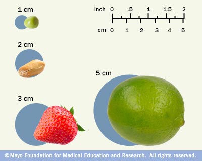
Tumor size - Mayo Clinic

View Image
View Image

Tumor-Induced Osteomalacia due to Hitherto Undiagnosed Subcutaneous  Phosphaturic Mesenchymal Tumor - AACE Clinical Case Reports
Tumor-Induced Osteomalacia due to Hitherto Undiagnosed Subcutaneous Phosphaturic Mesenchymal Tumor - AACE Clinical Case Reports